How Is a Brain Tumor Diagnosed? MRI, CT Scan and Biopsy Explained
Being told that a brain tumor may be present can be stressful for patients and their families. One of the most common questions is: How do doctors confirm whether a brain tumor is actually present?
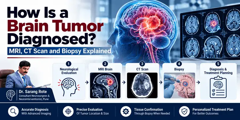
Brain tumor diagnosis usually involves more than one step. Doctors consider the patient's symptoms and medical history, perform a neurological examination, and may recommend imaging tests such as an MRI or CT scan. In some cases, a biopsy is needed to determine the exact type of tumor.
The diagnostic approach depends on the patient's symptoms, the location and appearance of the suspected tumor, and the information needed to plan treatment.
What Happens When a Brain Tumor Is Suspected?
A brain tumor may be suspected when a person develops neurological symptoms such as persistent headaches, seizures, changes in vision, weakness, numbness, speech difficulties, balance problems, memory changes, or changes in behaviour.
However, these symptoms can have many different causes and do not automatically mean that a person has a brain tumor. A doctor first evaluates the symptoms and performs an appropriate neurological examination before deciding which investigations are necessary.
The diagnostic process commonly includes:
- Medical history and symptom assessment
- Neurological examination
- MRI of the brain
- CT scan when appropriate
- Additional specialized imaging in selected cases
- Biopsy or surgical tissue sampling when required
Step 1: Medical History and Neurological Examination
The diagnosis usually begins with a detailed discussion about the patient's symptoms.
The doctor may ask:
- When did the symptoms begin?
- Are the symptoms getting worse?
- Have there been any seizures?
- Are there changes in vision or speech?
- Is there weakness or numbness?
- Have there been changes in memory, concentration, or behaviour?
- Is there a history of previous cancer or other neurological conditions?
A neurological examination may assess muscle strength, reflexes, coordination, balance, vision, eye movements, sensation, alertness, and other functions of the nervous system.
This initial evaluation helps the doctor understand which part of the nervous system may be affected and whether brain imaging is required.
Step 2: MRI Brain for Brain Tumor Diagnosis
What Is an MRI?
MRI stands for Magnetic Resonance Imaging. It uses a strong magnetic field and radio waves to create detailed images of the brain.
MRI is one of the most important imaging tests used when a brain tumor is suspected because it provides detailed information about soft tissues and can show the size, location, shape, and characteristics of an abnormal area.
MRI With Contrast
In many brain tumor evaluations, an MRI may be performed with a contrast material called gadolinium.
The contrast agent is given through a vein and can make certain abnormal areas more clearly visible on the scan. Contrast-enhanced MRI can help doctors better understand the appearance and extent of a suspected tumor.
Why Is MRI Important?
An MRI can help doctors determine:
- Where the abnormality is located
- How large it is
- Whether surrounding brain structures are affected
- Whether there is associated swelling or edema
- How the lesion relates to important brain structures
- How the lesion may have changed on follow-up imaging
MRI findings can also help doctors plan further investigations or treatment.
Step 3: CT Scan of the Brain
What Is a CT Scan?
A CT scan, or Computed Tomography scan, uses X-rays and computer processing to create detailed cross-sectional images of the brain.
CT can identify many brain abnormalities and may be particularly useful when a rapid evaluation is required. It can also provide useful information about bleeding, bone structures, calcification, and other findings.
MRI vs CT Scan for Brain Tumors
MRI and CT are not competing tests where one is always appropriate and the other is not. They provide different types of information and may be used at different stages of evaluation.
MRI generally provides better soft-tissue detail, making it particularly useful for evaluating suspected brain tumors.
CT is faster and more readily available in many emergency situations and can be particularly helpful when doctors need to quickly assess the brain or when MRI cannot be performed.
Your doctor decides which imaging test is appropriate based on the symptoms, clinical situation, and information needed.
Step 4: Specialized MRI Techniques
A standard MRI may sometimes be supplemented with specialized imaging techniques.
MR Spectroscopy
Magnetic Resonance Spectroscopy, or MRS, can provide information about certain chemical substances within brain tissue.
In selected cases, it may provide additional information about an abnormal area and can sometimes help doctors distinguish tumor tissue from other abnormalities.
MR Perfusion
MR perfusion evaluates blood flow through brain tissue.
Because some tumors have increased blood supply, perfusion information can provide additional details about a suspected tumor and may also help identify an appropriate area for biopsy in selected cases.
Functional MRI
Functional MRI, or fMRI, can help identify areas of the brain involved in important functions such as speech, movement, and sensation.
This information can be useful when a tumor is located close to an area responsible for an important neurological function and surgery is being considered.
Diffusion Tensor Imaging and Tractography
Diffusion tensor imaging and tractography can help visualize important white-matter pathways in the brain.
In selected patients, this information may contribute to surgical planning by showing the relationship between a tumor and important neural pathways.
Step 5: Is MRI Enough to Confirm a Brain Tumor?
An MRI can strongly suggest that an abnormal area is a brain tumor, and imaging can provide important information about its location and characteristics.
However, imaging alone does not always establish the exact tumor type.
Different conditions can sometimes have similar appearances on scans. When tissue confirmation is required, a biopsy or tissue obtained during surgery may be examined by a pathologist or neuropathologist.
This distinction is important because treatment depends on the specific type and characteristics of the tumor.
Step 6: What Is a Brain Tumor Biopsy?
A brain tumor biopsy involves removing a small sample of tissue so that it can be examined in a laboratory.
The tissue is examined under a microscope and may undergo additional laboratory and molecular testing to help identify the tumor type and other characteristics.
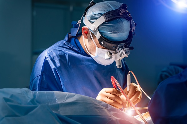
A biopsy may be performed separately or as part of surgery to remove the tumor.
Types of Brain Tumor Biopsy
Stereotactic Brain Biopsy
A stereotactic biopsy is a minimally invasive technique used to obtain tissue from a specific area of the brain.
Imaging guidance such as MRI or CT helps the neurosurgeon accurately guide the biopsy instrument to the targeted area.
This approach may be considered when a tumor is located deep within the brain, in an important functional area, or when removing the tumor completely through surgery may not be appropriate.
Surgical or Open Biopsy
If a tumor can be safely approached surgically, tissue may be obtained during an operation.
In some situations, instead of performing a separate biopsy, the neurosurgeon may remove as much of the tumor as can be safely achieved and send the removed tissue for pathological examination.
Why Is a Biopsy Important?
A biopsy can help determine:
- The exact type of tumor
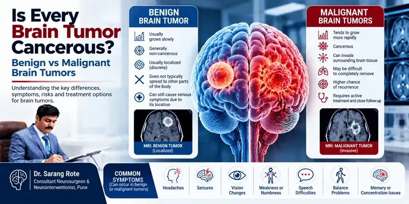
- Whether the tumor is benign or malignant
- The tumor's grade or biological characteristics
- Certain molecular or genetic characteristics
- Which treatment approaches may be appropriate
Modern brain tumor diagnosis may involve both microscopic examination and additional laboratory or molecular testing, depending on the suspected tumor type.
Does Every Brain Tumor Need a Biopsy?
No.
A biopsy is not appropriate or necessary in every situation.
Sometimes imaging characteristics and the clinical picture provide enough information to manage a lesion without immediately obtaining tissue. In other situations, biopsy may be considered too risky because of the tumor's location.
The decision depends on the individual case and the balance between the diagnostic information expected and the potential risks of the procedure.
What Happens After the Biopsy?
The tissue sample is sent to a pathology laboratory.
A pathologist or neuropathologist examines the sample and may perform additional tests to determine the tumor's characteristics.
The final pathology report helps the treating team understand what type of tumor is present and assists in planning the appropriate treatment.
Depending on the tumor, the pathology report may include information about the tumor type, grade, and relevant molecular findings.
Can a Brain Tumor Be Diagnosed Without Surgery?
In some situations, doctors can obtain substantial information from imaging studies without immediately performing surgery.
For certain lesions, the appearance on MRI or other imaging may strongly suggest a particular diagnosis, and the medical team may recommend observation or another treatment approach rather than immediate tissue sampling.
However, in many cases, tissue diagnosis remains important when the exact tumor type will influence treatment decisions.
What If the Tumor Is in a Difficult Location?
Some brain tumors are located close to areas responsible for critical functions such as speech, movement, vision, or other neurological functions.
In such cases, the diagnostic and treatment strategy must be carefully planned.
Advanced imaging techniques may help doctors understand the relationship between the tumor and important brain structures before deciding whether biopsy, surgery, observation, or another treatment approach is appropriate.
How Doctors Decide Which Test Is Needed
There is no single diagnostic test that is appropriate for every patient.
The diagnostic plan may depend on:
Symptoms
The type, duration, severity, and progression of symptoms can influence which tests are recommended.
Neurological Examination
The examination helps identify possible changes in brain or nerve function.
MRI Findings
MRI can provide detailed information about the location and characteristics of an abnormal area.
CT Findings
CT may provide important additional information, particularly in urgent situations or when evaluating bleeding, bone structures, or other abnormalities.
Tumor Location
The location of a suspected tumor can influence whether biopsy or surgery is considered safe.
Need for Tissue Diagnosis
If imaging cannot establish the diagnosis with sufficient certainty, tissue sampling may be necessary.
Treatment Planning
The tests selected may also depend on what information is needed to plan surgery or another form of treatment.
When Should You Consult a Neurosurgeon?
If brain imaging has identified a suspected tumor, consultation with a neurosurgeon can help you understand the findings and discuss the next steps.
A neurosurgical evaluation may be particularly important when imaging shows a mass, when neurological symptoms are persistent or progressive, or when biopsy or surgery is being considered.
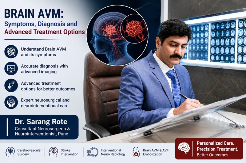
Dr. Sarang Rote's practice in Pune lists brain tumor surgery, brain tumors, skull-base neurosurgery, endoscopic neurosurgery, complex brain tumors, and brainstem tumors and other lesions among its areas of service and interest.
Why Choose Dr. Sarang Rote for Brain Tumor Evaluation?
Dr. Sarang Rote is a Consultant Neurosurgeon & Neurointerventionist in Pune. His website states that his areas of interest include skull-base tumors and other lesions, for which he offers microscopic and endoscopic skull-base neurosurgery, and it lists brain tumor surgery among his services.
His website also describes training in minimally invasive endoscopic brain and skull-base neurosurgery and lists experience and interests involving complex brain tumors, brainstem tumors, complex skull-base tumors, pituitary surgery, and tumor embolization.
For a patient with a suspected brain tumor, the diagnostic process should be individualized based on symptoms, imaging findings, tumor location, and the information required for treatment planning.
Frequently Asked Questions
Is MRI the best test for a brain tumor?
MRI is one of the most important imaging tests for evaluating suspected brain tumors because it provides detailed soft-tissue images of the brain. Specialized MRI techniques can provide additional information in selected cases.
Is a CT scan enough to diagnose a brain tumor?
A CT scan can identify many brain abnormalities and can be very useful in urgent situations. However, MRI generally provides greater soft-tissue detail for evaluating brain tumors. Depending on the situation, doctors may use both CT and MRI.
Does every brain tumor require a biopsy?
No. Some tumors can be managed based on their imaging appearance and clinical findings, while others require tissue confirmation. The decision depends on the individual case and the safety and usefulness of obtaining a biopsy.
Is a brain biopsy dangerous?
A brain biopsy is a medical procedure with potential risks, and the risks depend on factors such as the tumor's location and the method used. Doctors recommend biopsy when the expected diagnostic benefit justifies the procedure.
How long does a brain tumor diagnosis take?
The timeline varies. Imaging may provide important information quickly, while a final tissue diagnosis can take longer because the biopsy sample may require microscopic, immunohistochemical, and molecular testing.
Final Takeaway
Diagnosing a brain tumor is a step-by-step process rather than relying on a single test.
A doctor may begin with a detailed medical history and neurological examination, followed by imaging such as MRI or CT. If more information is needed, specialized imaging techniques may be used. In selected cases, a biopsy provides the tissue needed to establish the tumor type and guide treatment.
The right diagnostic approach depends on the patient's symptoms, imaging findings, tumor location, and overall clinical situation.
If you or a family member has been diagnosed with or is being evaluated for a brain tumor, consult a qualified neurosurgeon to understand the imaging findings and the appropriate next diagnostic step.















.jpeg)